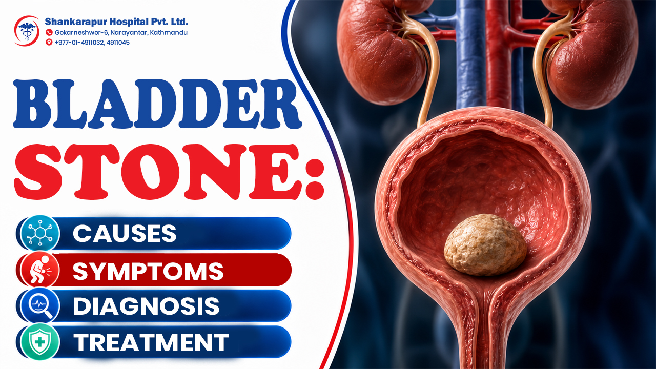
A bladder stone is a hardened mass of minerals that develops inside the urinary bladder. It most commonly forms when the bladder does not empty completely, allowing concentrated urine and mineral crystals to remain behind. Small stones may occasionally pass naturally, but many bladder stones require endoscopic fragmentation or surgical removal.
Treatment should not focus only on removing the stone. A urologist also needs to investigate why urine is remaining in the bladder, because an enlarged prostate, urinary blockage, nerve-related bladder dysfunction, infection or catheter may cause the stone to return.
This article explains bladder-stone symptoms, causes, diagnosis, treatment and removal options. It also clarifies the difference between a bladder stone and a kidney stone and identifies symptoms that require urgent medical attention.
What is a bladder stone?
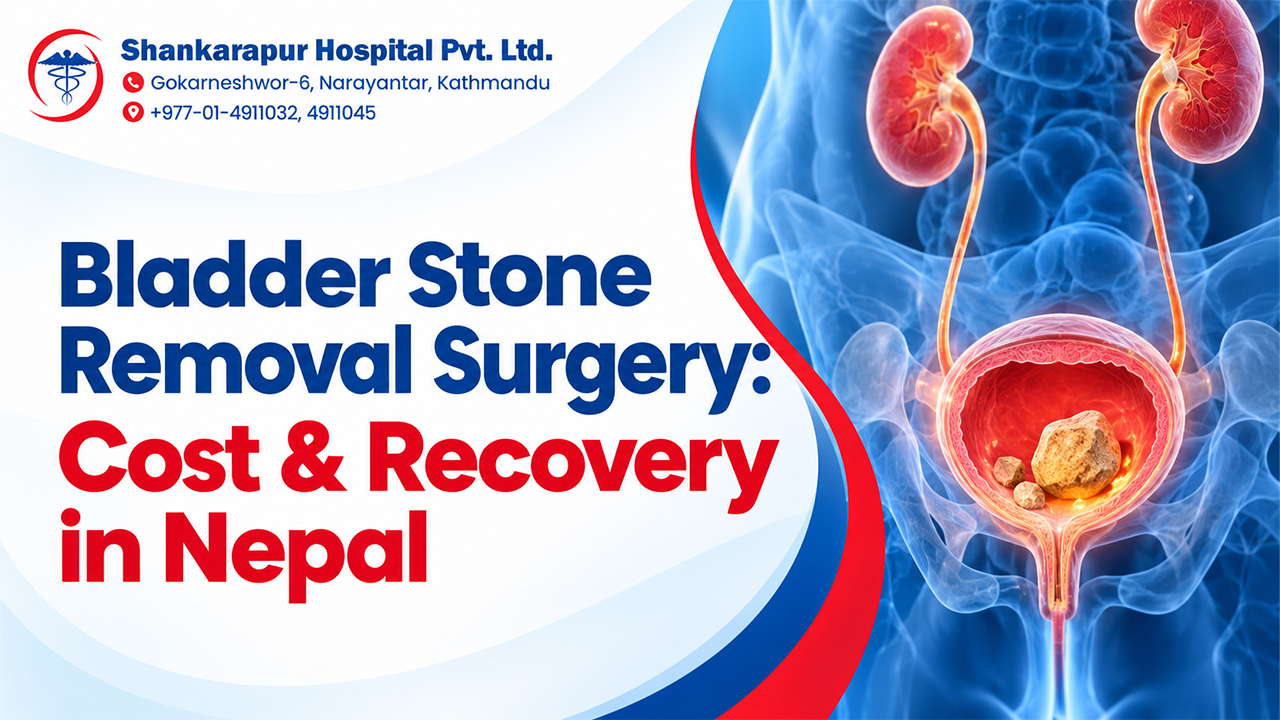
A bladder stone, also called a bladder calculus or vesical calculus, is a hard mineral mass located inside the urinary bladder. The bladder is a muscular organ in the pelvis that stores urine produced by the kidneys until it leaves through the urethra.
Stones can form when urine becomes concentrated and remains in the bladder for too long. Minerals in the retained urine begin to crystallize. The crystals may gradually combine, enlarge and harden into one or more stones.
Bladder stones are usually classified into three groups:
Primary bladder stones
Primary stones form inside the bladder without another obvious structural urinary problem. They are more commonly described in children living in settings where low fluid intake, recurrent diarrhoea and nutritional factors contribute to low urine volume or concentrated urine.
Secondary bladder stones
Secondary stones develop because another urinary condition interferes with bladder emptying or creates a surface on which crystals can collect. Common associations include an enlarged prostate, urethral narrowing, neurogenic bladder, chronic infection, bladder diverticula and long-term catheter use.
Migratory bladder stones
A migratory stone begins in a kidney or ureter and travels into the bladder. If it cannot leave through the urethra, it may remain in the bladder and become larger.
What causes bladder stones?
The most important cause of bladder-stone formation is incomplete bladder emptying. When urine remains after urination, minerals have more time to crystallize and combine.
The European Association of Urology reports that bladder outlet obstruction is the most common predisposing factor in adults and is associated with approximately 45%–79% of adult bladder-stone cases.
1. Enlarged prostate
An enlarged prostate, medically called benign prostatic hyperplasia or BPH, can press on the urethra and reduce urine flow. The bladder may then be unable to empty completely.
Urine left behind after urination is called post-void residual urine. Repeated retention creates an environment where crystals can settle and grow. Men with a weak stream, difficulty starting, stop-start urination, night-time urination or a persistent feeling of incomplete emptying may need assessment for prostate-related obstruction.
For a detailed explanation, see Shankarapur Hospital’s guide to enlarged prostate symptoms, diagnosis and treatment.
2. Urethral stricture or another urinary blockage
A urethral stricture is narrowing of the tube through which urine leaves the body. Scarring, previous procedures, injury, inflammation or another structural problem may restrict urine flow.
Any blockage between the bladder and the outside of the body can increase the amount of urine remaining after urination. A stone may then form or become trapped.
3. Neurogenic bladder
The bladder depends on coordinated nerve signals to store and release urine. Stroke, spinal-cord injury, Parkinson’s disease, diabetes, spinal conditions and other neurological problems may interfere with these signals.
Some people cannot contract the bladder strongly enough. Others cannot relax the urinary outlet at the correct time. Either problem can leave urine in the bladder and increase the risk of stones, infection or urinary retention.
4. Recurrent urinary infection
Some infections can contribute to stone formation, while an existing bladder stone can also encourage repeated infection. The stone may irritate the bladder lining and provide an area where bacteria remain.
Recurring burning urination, urgency, cloudy urine or infection that repeatedly returns should not be treated with leftover antibiotics. A clinician may need to check for a stone, obstruction, prostate problem or another structural cause.
Learn more about urinary tract infection symptoms and treatment.
5. Urinary catheters and foreign material
A catheter is a tube used to drain urine from the bladder. Long-term catheter use can increase bladder-stone risk because minerals may collect on the catheter or another foreign material.
People with permanent catheters, spinal-cord conditions or previous bladder reconstruction may need an individualized catheter-care and follow-up plan. They should not begin bladder irrigation or change catheter techniques without professional guidance.
6. Bladder diverticulum
A bladder diverticulum is a pouch in the bladder wall. Urine may remain inside the pouch even after the rest of the bladder empties.
Stagnant urine can support crystal formation, recurrent infection and stone growth. Treating the stone without assessing the diverticulum may leave the underlying problem unresolved.
7. A stone that migrated from the kidney
A kidney stone may travel through the ureter into the bladder. Many small stones leave the body during urination. However, a stone may remain if the bladder does not empty properly or if the urinary outlet is too narrow.
Once retained, it can act as a centre around which additional minerals collect.
8. Low urine volume and concentrated urine
Low fluid intake, heavy sweating, vomiting or diarrhoea can reduce urine volume. Concentrated urine contains a higher proportion of minerals relative to water.
Hydration matters, but dehydration is not the only explanation for most adult bladder stones. An adult who develops a bladder stone should also be evaluated for incomplete emptying, obstruction, infection or bladder dysfunction.
What are the symptoms of a bladder stone?
Bladder stones may cause no symptoms, especially when they are small. Symptoms usually develop when a stone irritates the bladder lining, moves near the bladder outlet or blocks urine flow.
Common bladder-stone symptoms include:
- Pain or burning while urinating
- Lower abdominal or suprapubic pain
- Frequent urination
- Sudden urinary urgency
- Difficulty starting urination
- A urine stream that stops and starts
- A weak urine stream
- Blood in urine
- Cloudy or unusually dark urine
- Recurrent urinary infections
- A feeling that the bladder has not emptied
Bladder-stone discomfort may become worse near the end of urination. Movement or physical activity can also increase symptoms for some patients. Recurrent UTIs may occasionally be the only noticeable sign.
Can a bladder stone cause blood in urine?
Yes. A stone can irritate or scrape the bladder lining and cause visible or microscopic blood in urine.
However, blood in urine has several possible causes, including infection, prostate disease, kidney disease, urinary-tract injury and bladder or kidney tumours. It should not automatically be blamed on a stone.
Read about other possible causes of blood in urine.
Can a bladder stone block urine?
Yes. A stone can move into the bladder outlet and prevent urine from entering the urethra.
Complete inability to pass urine is an emergency. It can cause severe lower abdominal pain and may require immediate drainage of the bladder as well as investigation of the obstruction.
Bladder stone vs kidney stone: What is the difference?
A bladder stone and a kidney stone are both urinary stones, but they form in different locations and often have different underlying causes.
| Feature | Bladder stone | Kidney stone |
| Where it forms | Inside the urinary bladder | Inside one or both kidneys |
| Common underlying issue | Incomplete bladder emptying or urinary obstruction | Mineral crystallization inside the kidney |
| Typical pain | Lower abdomen, bladder area, urethra or penis | Side, back, lower abdomen or groin |
| Urine flow | May be weak or stop and start | Usually changes when a stone reaches the lower urinary tract |
| Common associated condition | Enlarged prostate, neurogenic bladder, catheter or stricture | Low urine volume, metabolic factors, diet, family history or certain medical conditions |
| Usual treatment | Bladder-based endoscopic fragmentation or removal | Observation, ureteroscopy, shock-wave treatment, RIRS or PCNL depending on location and size |
| Key recurrence strategy | Correct the emptying problem | Identify metabolic, dietary and urine-volume risks |
Kidney stones form in the kidney and may cause severe wave-like pain when they move into a ureter. Bladder stones tend to cause lower urinary symptoms, such as painful urination, stop-start flow, frequency and incomplete emptying.
A kidney stone can become a bladder stone if it reaches the bladder but cannot leave.
Readers concerned about stones in the kidney or ureter can review kidney-stone treatment options in Nepal.
How are bladder stones diagnosed?
A urologist diagnoses a bladder stone by combining the symptom history, physical examination, urine tests and appropriate imaging. The assessment should also identify why the stone formed.
Medical history and examination
The clinician may ask about:
- Pain, burning or blood during urination
- Frequency, urgency or night-time urination
- Weak or interrupted urine flow
- Urinary retention
- Previous kidney stones
- Prostate symptoms
- Urinary infections
- Catheter use
- Neurological conditions
- Previous urinary surgery
- Medicines and supplements
The examination may include checking the lower abdomen for bladder distension. In men, the clinician may evaluate whether prostate enlargement could be contributing to obstruction.
Urine test and urine culture
Urinalysis can look for blood, infection, crystals and urine acidity. A urine culture may be required when infection is suspected.
Urine testing cannot always prove that a stone is present, but it can identify complications or clues about the stone’s composition.
Ultrasound
The European Association of Urology recommends ultrasound as first-line imaging for symptoms suggestive of a bladder stone. Ultrasound does not use ionizing radiation and can also help evaluate post-void residual urine.
CT scan
CT can detect small stones and show their location, size and number. It can also assess the kidneys and ureters when an upper urinary-tract stone is possible.
CT is not required for every patient. The clinician considers symptom severity, ultrasound findings, medical history, radiation exposure and whether another condition needs to be excluded.
Cystoscopy
During cystoscopy, a thin camera is passed through the urethra into the bladder. It allows the urologist to look directly for a stone and examine the bladder lining, urethra and outlet.
Cystoscopy may be considered when symptoms persist, imaging is unclear or another bladder condition needs to be ruled out.
Uroflowmetry and post-void residual measurement
Uroflowmetry measures the speed and pattern of urine flow. Post-void residual measurement checks how much urine remains after urination.
These tests are important because removing a stone without understanding a weak flow or incomplete emptying may not prevent recurrence. EAU guidance recommends investigating the cause with urine testing, flow assessment, residual urine measurement and metabolic or stone analysis where appropriate.
Shankarapur Hospital’s diagnostic and imaging services publicly list ultrasound and CT support. The urologist determines which tests are appropriate for each patient.
Can a bladder stone pass naturally?
A small bladder stone that migrated from the upper urinary tract may occasionally pass during urination, particularly when there is no bladder outlet obstruction, bladder dysfunction or long-term catheter.
However, most primary and secondary bladder stones are unlikely to pass naturally. They commonly form because the bladder is not emptying properly, and the same problem can prevent the stone from leaving.
Will drinking more water remove a bladder stone?
Drinking water may help a very small stone pass and may reduce urine concentration. It does not reliably remove a stone when the bladder outlet is blocked or the bladder muscle cannot empty effectively.
People with heart failure, kidney disease or another condition requiring fluid restriction should ask their clinician how much fluid is appropriate.
Can medicine dissolve a bladder stone?
Most bladder stones cannot be quickly dissolved with medicine.
Some confirmed uric-acid stones may be treated with medically supervised urinary alkalinization. This requires accurate identification of the likely stone type, regular urine-pH monitoring and clinician-directed treatment. It should not be attempted with household remedies or unmonitored supplements.
Bladder-stone treatment and removal options
Bladder-stone treatment depends on:
- Stone size, number and hardness
- Symptoms
- Infection
- Ability to pass urine
- Bladder function
- Prostate size
- Urethral narrowing
- Previous urinary surgery
- Catheter use
- Age and general health
- Anaesthesia considerations
Many symptomatic bladder stones require active removal. Minimally invasive endoscopic procedures are generally preferred when suitable.
1. Observation
Observation may be reasonable for a small, asymptomatic migratory stone when the urologist believes it can pass and there is no urinary obstruction, bladder dysfunction, infection or long-term catheter.
Monitoring should include clear instructions about worsening pain, urinary blockage, fever or blood in urine.
2. Transurethral cystolithotripsy or cystolitholapaxy
This is a common minimally invasive bladder-stone removal method.
A small endoscopic instrument is passed through the urethra into the bladder. The urologist sees the stone directly and breaks it into smaller fragments using laser, ultrasonic, pneumatic or another fragmentation technology. The fragments are then removed or washed from the bladder.
The terms cystolithotripsy and cystolitholapaxy are sometimes used in overlapping ways. Both generally describe endoscopic fragmentation and removal of a bladder stone.
EAU guidance recommends transurethral cystolithotripsy for adults where possible.
3. Percutaneous cystolithotripsy
In a percutaneous procedure, the urologist reaches the bladder through a small opening in the lower abdomen rather than through the urethra.
This approach may be considered when the stone is difficult to manage through the urethra, when urethral access carries additional risk or when the patient’s anatomy or previous surgery makes another route preferable.
4. Open cystolithotomy
Open surgery may be considered for a very large or exceptionally hard stone that cannot be safely or efficiently removed with an endoscopic procedure.
The stone is removed through an incision into the bladder. Recovery and hospital requirements are generally greater than with a minimally invasive procedure, so the urologist balances stone size, anatomy and patient health before recommending it.
5. Shock-wave treatment
Shock-wave lithotripsy uses externally delivered energy to fragment a stone.
It can be an alternative in selected cases, but European guidance indicates that stone-free rates are generally lower than with endoscopic or open procedures for bladder stones. The urologist considers stone size, composition, equipment availability and the likelihood that fragments can leave the bladder.
Bladder-stone treatment comparison
| Treatment | May be suitable for | Main advantage | Important limitation |
| Observation | Selected small, asymptomatic migratory stones | Avoids an unnecessary procedure | Many bladder stones will not pass |
| Medically supervised dissolution | Selected confirmed uric-acid stones | May avoid surgery | Works only for specific stones and needs monitoring |
| Transurethral cystolithotripsy | Many adult bladder stones | No external incision; direct fragmentation and removal | Urethral access must be suitable |
| Percutaneous treatment | Stones difficult to remove through the urethra | Direct access to the bladder | Requires a small abdominal access tract |
| Open cystolithotomy | Very large or difficult stones | Allows removal of a large stone | More invasive and generally requires greater recovery |
| Shock-wave treatment | Selected patients | No instrument may need to enter the bladder | Fragments still need to pass; lower stone-free rates in some cases |
The preferred procedure is not determined by size alone. Urinary obstruction, urethral anatomy, infection, bladder function, anaesthesia safety and whether another operation is needed may change the decision.
Why treating the underlying cause matters
Removing the stone solves the immediate problem, but it may not solve the reason the stone formed.
Before or during treatment, the urologist may investigate:
- Enlarged prostate
- Urethral stricture
- Weak bladder contraction
- Neurogenic bladder
- Bladder diverticulum
- Chronic infection
- Catheter-related factors
- A stone migrating from the kidney
- Metabolic or urine-composition abnormalities
Enlarged prostate and bladder stones
When prostate enlargement causes significant obstruction, the urologist may recommend treating the obstruction as well as removing the stone.
Treatment might be performed during the same admission or in a planned sequence. The decision depends on prostate findings, infection, bleeding risk, anaesthesia considerations and the patient’s priorities.
Shankarapur Hospital’s Urology Department lists medical and surgical urology care, endoscopic and laser stone procedures and prostate-enlargement treatment through TURP. Specific suitability and current availability should be confirmed during consultation.
Infection and bladder stones
When a urine infection is present, treatment may include a prescribed antibiotic based on clinical findings and, when appropriate, urine-culture results.
Antibiotics do not remove the stone. If the stone or urinary blockage is allowing infection to return, structural treatment may also be necessary.
Catheter and neurogenic bladder
Patients who depend on urinary catheters or intermittent catheterization may need a personalized plan covering catheter technique, bladder emptying, infection prevention and follow-up.
Bladder irrigation is recommended only for specific high-risk groups and must be guided by a qualified professional. It is not a general home treatment for every person with a bladder stone.
What affects bladder-stone treatment cost in Nepal?
A reliable treatment quotation usually requires examination and imaging. Costs may differ according to:
- Consultation and diagnostic tests
- Ultrasound, CT or cystoscopy requirements
- Number and size of stones
- Endoscopic, percutaneous or open technique
- Laser or other fragmentation technology
- Anaesthesia
- Catheter requirements
- Infection treatment
- Hospital stay
- Stone analysis
- Treatment of an enlarged prostate or stricture
- Follow-up visits and repeat imaging
A very low procedure-only quotation may not include imaging, anaesthesia, medicines, hospital charges or treatment of the underlying obstruction. Patients should ask for a written explanation of what is and is not included.
Recovery after bladder-stone removal
Recovery varies by procedure and by whether another urinary problem is treated at the same time.
Temporary symptoms may include:
- Burning while urinating
- Mild blood staining in urine
- Urinary urgency
- Bladder discomfort
- Temporary catheter use
The treating team should explain expected symptoms, medicines, activity restrictions, catheter care and warning signs before discharge.
Patients should seek advice promptly if they develop increasing pain, inability to urinate, fever, chills, heavy bleeding, clots, repeated vomiting or worsening weakness.
Can bladder stones come back?
Yes. Recurrence is more likely when the cause of incomplete bladder emptying remains untreated.
The most important prevention step is therefore not a single food restriction or supplement. It is identifying and managing the person’s individual risk factors.
Practical prevention measures
- Treat enlarged prostate or urinary obstruction when clinically appropriate.
- Follow the recommended catheter or bladder-emptying plan.
- Treat confirmed urinary infections correctly.
- Drink an appropriate amount of fluid unless medically restricted.
- Avoid repeatedly delaying urination.
- Attend follow-up when residual urine or bladder dysfunction is present.
- Ask whether the removed stone should be analysed.
- Discuss metabolic assessment if stones recur or if kidney stones are also present.
There is limited evidence for a universal diet that prevents every type of bladder stone. Dietary recommendations should reflect stone composition, kidney function and other medical conditions rather than a generic prohibited-food list.
Common mistakes to avoid
Assuming every urinary stone is a kidney stone
Pain, investigation and treatment differ depending on whether the stone is in the kidney, ureter or bladder.
Relying only on water
Hydration may help a very small stone, but it does not correct prostate obstruction, urethral narrowing or weak bladder contraction.
Treating repeated infections without checking the urinary tract
Recurring infection can be a sign of a retained stone, obstruction or another structural problem.
Removing the stone but ignoring the cause
A successful procedure does not guarantee that another stone will not form if urine continues to remain in the bladder.
Using alkalizing products without identifying the stone
Only selected uric-acid stones may respond to urinary alkalinization. Incorrect use may be ineffective or unsafe.
Ignoring visible blood in urine
A stone can cause bleeding, but so can other urinary conditions. Visible blood should receive appropriate assessment.
When should bladder-stone symptoms be treated urgently?
Seek urgent medical care when urinary symptoms include:
- Complete inability to pass urine
- Severe or rapidly worsening lower abdominal pain
- Fever or chills with painful urination
- Repeated vomiting or inability to drink
- Heavy visible bleeding or blood clots
- Confusion, faintness or marked weakness
- Severe symptoms in a child, pregnant patient or person with major kidney disease
Untreated bladder stones may cause chronic urinary problems, recurrent infection or blockage of urine flow. A bladder stone is not usually immediately life-threatening, but urinary obstruction or infection can become serious and should not be delayed.
Shankarapur Hospital states that its Emergency & Critical Care Department operates 24 hours a day.
Choosing urology care in Kathmandu
A patient does not need to choose a hospital based on an unsupported “best hospital” claim. A more practical approach is to check whether the provider can investigate the stone, determine why it formed and offer an appropriate treatment pathway.
Questions to ask include:
- Is the stone definitely in the bladder?
- How many stones are present, and how large are they?
- Is urine remaining after I urinate?
- Could an enlarged prostate or stricture be causing obstruction?
- Is there a urine infection?
- Which removal method is recommended and why?
- Will the underlying cause be treated at the same time?
- Will the stone be sent for analysis?
- What are the likely risks and recovery requirements?
- What costs are included in the treatment estimate?
- What follow-up is needed to reduce recurrence?
Shankarapur Hospital publicly lists a Urology Department, diagnostic and imaging services, specialist consultations, inpatient wards and 24/7 emergency support. It also lists Dr. Wesh Ansari, Urology & Kidney Transplant Surgeon.
People experiencing persistent bladder-stone symptoms can request a urology appointment for an individualized assessment.
Key takeaways
- A bladder stone is a hard mineral mass inside the urinary bladder.
- Many adult bladder stones form because the bladder does not empty completely.
- Enlarged prostate, urinary blockage, nerve-related bladder dysfunction, infection and catheters are important causes.
- Symptoms can include painful urination, lower abdominal pain, frequent urination, blood in urine and a stop-start stream.
- Bladder stones and kidney stones form in different locations and may require different procedures.
- Ultrasound is commonly used as first-line imaging, with CT or cystoscopy used when necessary.
- Small migratory stones may occasionally pass, but many bladder stones need endoscopic removal.
- Successful treatment should address both the stone and its underlying cause.
- Inability to urinate, fever with urinary symptoms or severe worsening pain requires urgent care.
FAQs
What are the symptoms of a bladder stone?
Bladder-stone symptoms can include lower abdominal pain, burning or pain during urination, frequent urination, urgency, a weak or interrupted urine stream, blood in urine and cloudy or dark urine. Some people experience repeated urinary infections, while small stones may cause no symptoms. Symptoms cannot confirm a stone without medical evaluation and appropriate testing.
How do I get rid of bladder stones?
A very small stone may occasionally pass naturally, but most symptomatic bladder stones require removal by a urologist. A common treatment is transurethral cystolithotripsy or cystolitholapaxy, in which a scope enters through the urethra and a laser, ultrasound or another device breaks the stone into removable fragments. Large stones may require percutaneous or open surgery.
Is a bladder stone life-threatening?
A bladder stone is not usually immediately life-threatening. However, it can block urine flow, cause recurrent infection, produce bleeding or contribute to long-term bladder problems. Complete inability to urinate, fever or chills with urinary symptoms, severe worsening pain or marked illness requires urgent medical assessment.
What is it like to pass a bladder stone?
A very small bladder stone may pass without being noticed. When symptoms occur, passing it may cause burning, lower abdominal or urethral discomfort, urgency or a temporary interruption in the urine stream. A person may notice a small hard particle after urination. Severe pain, bleeding or difficulty passing urine should not be managed only at home.
Can a bladder stone go away?
A small migratory bladder stone may pass on its own when there is no urinary obstruction or bladder dysfunction. Primary and secondary stones are less likely to pass because they often form in a bladder that does not empty properly. Selected uric-acid stones may sometimes be dissolved with medically supervised urinary alkalinization, but most stones cannot be dissolved with home remedies.
What are five warning signs of a bladder stone or another bladder problem?
Five important warning signs are painful urination, visible blood in urine, frequent or urgent urination, a weak or stop-start urine stream and persistent lower abdominal pain. Recurrent UTIs or inability to empty the bladder are also concerning. These symptoms are not specific to stones and may occur with infection, prostate problems or other urinary conditions.
Can bladder stones cause repeated urinary infections?
Yes. A bladder stone can irritate the bladder and create conditions where infection returns. Recurrent infection may also be a sign that the bladder is not emptying or that a catheter, prostate obstruction or another urinary problem is present. Treating infection alone may not prevent recurrence if the stone or underlying cause remains.
Can women and children get bladder stones?
Yes. Although bladder stones are more common in older men, women and children can also develop them. Women may have urinary obstruction, bladder dysfunction, infection or foreign material. In some children, low urine volume, recurrent diarrhoea and nutritional factors may contribute. A child with pain, urinary retention, blood in urine or repeated infection needs professional assessment.